


American Laboratory Features Microtrace Scientist’s Photomicrograph
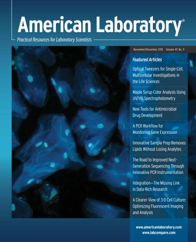
The cover of this month’s American Laboratory magazine features a photomicrograph taken by Microtrace scientist Katie White. Katie collected this image by fluorescence microscopy under ultraviolet excitation at 40x magnification.
The photograph shows epithelial cells from a cheek that was treated with DAPI, a fluorescent stain that binds to nucleic acids, such as the DNA found in cells. Although the thin cell membranes also fluoresce due to DNA, the rounded nuclei of the cells appear particularly bright since they contain most of the cell’s genetic material. This photograph illustrates both the microscopic morphology of our cheek cells and the bright blue fluorescence that they exhibit in DAPI stain.
How May We Help You?
Contact usto discuss your project in more detail.