


Fluorescence Microscopy
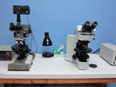
A fluorescence microscope uses an ultraviolet light source (rather than visible light) to illuminate a specimen. The ultraviolet radiation is absorbed by certain compounds, which is then re-emitted in the visible wavelength. This phenomenon, known as luminescence (fluorescence or phosphorescence, depending on the timescale for re-emission), depends upon the property of the compound being probed. While we commonly use this tool to examine biological materials (unknown or suspected cells) using fluorescent stains (e.g., DAPI), the technique is also particularly useful in particle analysis to identify the presence of optical brighteners, dyes and other autofluorescing substances that are encountered. Microtrace maintains two different Olympus microscopes (BH and BX-51) configured for fluorescence microscopy (one with a traditional mercury burner, the other with an X-Cite liquid light guide) as well as a microspectrofluoimeter configured with xenon (Xe) and mercury (Hg) illuminators for quantitative spectroscopic characterization of fluorescence on microscopic particles.
How May We Help You?
Contact usto discuss your project in more detail.